Categories
Blogs

Is a Liver Infection Serious?

Is a Liver Infection Serious?

The liver is a large organ around the size of a football that functions as a storage organ. It’s located on the right side of your abdomen, straight below your rib cage. The liver is important for the digestion of food and eliminating waste from the body.

Liver disease may be inherited genetically. Viruses, excessive alcohol use, and obesity are a few factors that may cause liver damage.

Liver damage may further progress into liver scarring (cirrhosis), leading to liver failure, a life-threatening condition. On the other hand, early diagnosis and treatment may enable the liver to heal.

What does infection in the liver mean?


When disease-causing agents (bacteria, parasites and viruses) infect your liver cells, inflammation is triggered. This impairs the normal functioning of the liver. As a result, symptoms like tiredness, itchy skin, dark urine, chronic fatigue, pale stool, diarrhoea, and loss of appetite may show up.

The diseases causing pathogens can enter the liver through blood, contaminated water or food, or contact with an infected person. The most common form of liver infection includes Hepatitis A, B, and C caused by the hepatitis group of viruses.

In addition to jaundice, several of these symptoms might signal various other problems, from stomach bleeding to heart failure to Wilson’s disease. Inheritance of an abnormal gene from one or both of your parents can cause a build-up of various substances in your liver, leading to liver damage.

For example, in Wilson’s Disease, this hereditary ailment develops when copper builds up in your body, harming your brain and liver. A lack of specific symptoms may make it difficult to recognise liver failure since toxins can build up in your body and brain while your liver is out of action, leading to additional health issues, including liver cancer.

What virus causes liver infection?


Hepatotropic viruses infect and multiply in the liver, causing illness. This group of viruses includes the hepatitis A, B, C, and E viruses. Hepatitis and liver damage occurs because of the liver’s immunological response to a virus in any of these illnesses.

The liver is prone to further damage because of greater host infection with viruses that predominantly attack other organs, notably the upper respiratory tract. Herpes viruses are an example of this phenomenon.

What happens if the liver is infected?

Some types of liver disease may raise your risk of developing liver cancer. If you do not get your liver checked properly, it will worsen. Cirrhosis (liver scarring) is a condition that arises when this happens.

A damaged liver will ultimately run out of healthy tissue and not perform its essential functions. If untreated, liver disease may progress to organ failure.

Read More – How Can Stomach Cancer Be Prevented?

Is liver infection contagious?


The liver infection can’t be spread by casual contact with an infected person. However, getting hepatitis by blood, faeces, or sexual contact is possible. It is one of the most common causes of liver illness. Infectious diseases, such as chickenpox, can cause liver damage, although rare.

Is a liver infection fatal?


Bacterial infection is a common and often fatal consequence of liver illness. It may cause mortality directly or indirectly by precipitating gastrointestinal bleeding, hepatic encephalopathy, or renal failure.

How long does a liver infection last?


Viral hepatitis like hepatitis A and E virus last for a few days to a few weeks. Moreover, the patient recovers after a supportive treatment and develops a lifelong immunity towards those viruses.

However, hepatitis B and C can behave like acute viral illness, but in some patients they might become lifelong carriers. This causes chronic liver damage leading to chronic liver disease like cirrhosis, or malaganices.

Hence, it is crucial to consult a doctor if you develop a hepatitis infection.

Can liver infections be cured?


Some liver conditions may be cured since a cure entails restoring good health and recovery. However, some liver illnesses cannot be treated, but the symptoms may be managed. For example, the alcohol-related liver impairment may be effectively managed with proper diet and exercise, and your liver may ultimately recover.

On the other hand, your liver may fail if the circumstances are sufficiently severe. Surgery may help with certain disorders. People with serious illnesses may be candidates for a liver transplant, which might repair the condition; however, recovery time is lengthy.

Conclusion


It is possible to develop liver disease as a consequence of an infection, an inherited illness, cancer, or an excess of poisonous chemicals. Healthcare practitioners may properly treat many forms of liver disease with medicine or by adopting a healthier lifestyle. If you have serious liver disease, a liver transplant may be an option to help you regain your health and live longer.

Contact our experts today to know more about liver diseases and its cure.

Categories
Blogs

How Can Stomach Cancer Be Prevented

How Can Stomach Cancer Be Prevented?

Stomach cancer is characterized by the proliferation of malignant cells in the abdominal wall. Most individuals don’t display any symptoms in the early stages of gastric cancer, also known as gastric adenocarcinoma. It is difficult to accurately diagnose stomach cancer since it is so uncommon when matched to other forms. Because stomach cancer typically has no early signs, it might go undetected for a long time until it is discovered. This may further complicate the treatment process.

What causes most stomach cancer?


An abnormal genetic mutation in the stomach’s lining is the primary reason for stomach cancer. This mutation allows the stomach cells to develop fast and ultimately form tumours. The following are potential causes that might raise a person’s likelihood of developing stomach cancer:

  • Family history of cancer
  • Smoking Cigarettes
  • Overweight
  • Inherited mutations in certain genes may increase the risk of stomach cancer. But only a tiny fraction of stomach cancers are caused by this.
  • Infections from Helicobacter pylori bacteria can increase the risk of stomach cancer.
  • A high-sodium diet 
  • And a diet that is low in fruit and veggies.

Genetic mutations after birth can also contribute to stomach cancer. Certain risk factors, like cigarette smoking before or during pregnancy, can develop these inherited mutations. Alternatively, certain gene modifications can also occur with no external trigger and cause cancer.

Stomach Cancer, Diet, and nutrition


You can fight any illness if you follow the right nutrition. We’ve compiled a list of certain foods that may aid in preventing and treating cancer and foods that can reduce the risk of stomach cancer.

  • Consume foods high in quercetin – Consume lots of onions, citrus fruits, apples, cherries, and broccoli. These foods have high levels of quercetin, which may help prevent this cancer from developing.
  • Make use of Turmeric – Turmeric contains a chemical compound known as curcumin. It has potent anti-cancer capabilities, and it causes cancer cells to activate their own self-destruct mechanisms.
  • Allicin-rich foods – In allicin content, garlic is the most abundant supply available and is quite efficient against such a malignancy. The allicin found in onions is also a potent source of vitamin C.
  • Vitamin A, C, and E-rich foods are available – Vitamin E can be found in spinach and almonds. In contrast, vitamin C can be found in abundance in citrus fruits and tomatoes, whereas vitamin A can be found in abundant amounts in carrots and potatoes.
  • Eliminate foods that have been smoked, heavily salted, or smoked
  • Minimize intake of animal fats.
  • Restrict consumption of alcoholic beverages.

Tips For Preventing Stomach Cancer


Even though stomach cancer is the 4th most frequent disease globally, the majority of instances have decreased significantly over the last few decades. Unfortunately, there is no certainty that you will be able to avoid it, but there are several things you could do to reduce your risks of getting it. Here are some tips to prevent the risk of cancer.

  • Reduce your intake of smoked foods – foods like pickles or smoked food items have preservatives and high salt content. Large levels of salt and additives may cause damage to the wall of the stomach, increasing your risk of developing stomach cancer. As a result, restrict your intake of smoked and pickled meals, as well as salted meats and seafood.
  • Take, for example, genetic testing – Is stomach cancer something that runs in your relatives? In certain cases, a genetic test may determine if you have particular genes (CDH1 gene) to make you more susceptible to stomach cancer.
  • Aspirin should be used with caution – To relieve pain, temperature, and inflammation, you might take ibuprofen, aspirin, or other over-the-counter medications similar to it. This treatment may also reduce your risk of developing tummy and colorectal cancers. However, you should avoid using such drugs to avoid stomach cancer since they can induce internal bleeding. Your physician will be able to tell you what is best for you.

Conclusion


The incidence of stomach cancer has steadily decreased during the last century. It appears as though the number of reported cases and fatalities from stomach disease around the globe has been stable in recent years. Furthermore, there has been a steady decline in the incidence of stomach cancer in the region nearest to the duodenum. Nevertheless, cancer incidence at the oesophageal-gastric junction is on the rise.

To know more about different ways to prevent and reduce your risks of stomach cancer, contact our experts today.

Categories
Blogs

Choosing the right oncosurgeon

Choosing the right oncosurgeon

 Fear, anger, disbelief, rejection… these are just some of the emotions that a person goes through when they hear a diagnosis of cancer.

This is true for almost all cancer patients, but equally true is the fact that they all come to terms with it… some sooner, some a bit later. And the sooner you make peace with the reality, the faster you can start taking proactive steps to wage a battle against cancer.

There are different cancers and different stages which call for different treatment modalities. Some may require chemotherapy, some radiation therapy, some may require surgery, and some may require a combination of two or more of the above interventions.

In this article we are focusing primarily on choosing an oncosurgeon, (if you have been advised surgery) since getting operated is always a scary thought and choosing a surgeon is the hardest part. unless you have full faith and confidence in the competence and credibility of your oncosurgeon, you will always have second thoughts. So choose wisely.

Let us try and understand how we can make this choice, a well informed and a well researched one:

  1. Referring Physician: This would most probably be your family doctor or physician. They are the ones who are most familiar with your medical history and also the ones you have the most faith in. They have, in all probability, referred you to an oncosurgeon, who is best suited to treat your particular type of cancer. They would also be the ones who would be closely involved with you and the operating doctor throughout the course of your treatment. So your best bet would be to consult with the specialist, they have referred to.
  2. Second opinion: Whatever your stage of cancer, there is always time for a second opinion. A second opinion is important as it helps you to compare treatment protocols. Check whether the recommendations of the 2 doctors match each other or there is a difference of opinion. Also helps to compare other things such as facilities offered, choice of hospital, techniques of surgery, cost of treatment, etc. It helps you make a more informed decision. Remember, in the end its your choice.
  3. Background search: It wouldn’t hurt to do some background research about the doctor you have been referred to on your own, either on the internet or through your friends and family who have had some direct or indirect experience about that cancer specialist. This research should typically be focused on:
    1. The credentials of the oncosurgeon. What is his/her degree? Is it from a reputed institution and/or national or state board? Have they done any specialisation in the field of oncosurgery?
    2. Do they focus on any specific organ or do they perform multi-organ surgeries.
    3. Are there any advanced techniques which they are skilled in such as robotic surgery, minimally invasive surgery, or any technique which they have patented or pioneered in?
    4. Have they received any special training in treating your specific type of cancer?
    5. What is their ability to handle complicated cases as well as what is the percentage of complications expected and encountered by them specific to your type of cancer?If you don’t find answers to any of the questioned mentioned above or have any other questions in mind, you can always ask them to the doctor, without being embarrassed about it. Its your right to know.
  4.  Experience of the oncosurgeon: experience both in terms of number of years in practice as well as the number of surgeries performed. A doctor who has more experience in operating on a certain type or types of cancers, has more experience in dealing with intraoperative complications and resolving them and consequently has more likelihood of giving better results.
  5. Personality: Many would argue that the personality of a doctor or their approach towards the patient has nothing to do with his/her skills in the operation theater, but there is an important component of any doctor-patient relationship and that is “the comfort level.” Unless you are comfortable sharing your doubts and queries with the doctor and get the feeling that the doctor is genuinely interested in hearing your problems and always has your best interest at their heart, you would not like to get operated from such a doctor, however competent or reputed. So the most important thing to look for in a doctor is approachability. He/she should have a pleasant personality, make you feel important, be willing to clear all your doubts, however silly, and most importantly take your preferences and wishes into consideration.
  6. Additional facilities: A cancer surgery is not a walk-in, walk- out procedure. The entire process may take anywhere from a few days to a few weeks. So, many other factors should be taken into consideration while selecting an oncosurgeon:
    1. Is he/she going to operate in a multispeciality hospital with state of the art latest equipments and a large ICU setup with round the clock monitoring
    2. what are the types of accommodation facilities at the hospital for you and your attendant, relative or friend.
    3.  Does he/she have a team of doctors which can handle other related aspects of the disease such as, your medicine or surgical side effects like pain, nausea, vomiting, infections, etc.
    4. Does he/she have a team of support staff such as an on-call secretary through whom you can be in constant touch with your surgeon, a billing assistant, a mental health counsellor, a follow- up counsellor, a diet counsellor, a physical therapist etc.
    5. what are the modes of payment accepted by your surgeon and the hospital. Do they have cashless mediclaim facility, do they have medical loan facility, do they have installment facilities etc.
  7. Patient testimonials and reviews: Nothing puts a troubled mind to ease more than knowing that someone else has gone through what you are going through right now and come out victorious. So reading testimonials of other patients who have been treated by the doctor of your choice is the best way to judge the competence of your doctor. You should be able to find patient testimonials at your oncosurgeons office either in written form or as a video. Alternatively you can ask for contact number of patients who are willing to share their story. You would be surprised, that most of the times, you will find answers to questions you had never even thought of. You can also read reviews on social media and the internet, but beware, along with genuine reviews, there are a lot of negative comments posted by people with mala fide interests. Use your better judgement.
  8. Be informed, but not influenced: Information is never a bad thing. But knowing the difference between the right and the wrong kind of information is of paramount importance, especially when it relates to something as serious as cancer. For every 1 piece of well researched, well documented, scientifically and  medically proven treatment modality, there are hundreds of baseless claims, unresearched, unproven alternative treatments offered with the promise of a sureshot cure. Please don’t believe in it. Not only do they give you false hope, they end up creating more confusion and chaos, which delays your treatment and reduces your chances of a quick recovery. It is a good idea to learn more about your disease, the kind of treatment options available, the latest developments and research in the field. It helps you frame your questions, more accurately for your doctor. And if you have read or heard about any such alternative treatment option, place it in front your oncosurgeon during your consultation. They will be more than willing to clear your doubts.  Finally, just like there are no guarantees in life, so too, no cancer surgeon can guarantee that you would be totally cured or that there wont be any complications during or after your surgery. But if you have taken the efforts to do due-diligence while selecting your surgeon, it will stack the odds highly in your favour.
Categories
Blogs

CAR T cells

CAR T cells

 Chimeric antigen receptor (CAR) T cell therapy is a new form of cancer therapy and is showing a lot of promise.

T cells are a part of body’s immune system. They are a type of white blood cells. T cells have proteins on their surface which act as receptors. Any foreign substance that enter the body like bacteria and virus also have proteins on their surface. Whenever T cell receptors come across the protein antigen of the foreign substance, the latch onto it and destroy the foreign substance. But Not every T cell receptor can bind to every protein antigen since both the receptors as well as antigen on foreign bodies are differently shaped. Only those that fit their receptors can be destroyed.

This forms the basis of CAR T cell therapy in cancer therapy. Since cancer cells also have antigens and can bind to T cells receptors but it is rarely a perfect fit, scientists have developed a novel method of help the T cells bind more accurately to a particular type of cancer cell.

The process:

  1. Firstly patients are evaluated thoroughly via a series of tests to assess if CAR T cell therapy would be useful in their case or not

  2. Secondly blood is drawn patients into a machine where white blood cells are removed along with the T cells and remaining blood is sent back into the patients body.

  3.  Next, T cells are separated from the rest of the white cells. They are sent to the laboratory where Chimeric antigen receptors CARs are added to the T cells, creating CAR T cells which can recognize antigen on the targeted tumor or cancerous cells.

  4. These reengineered T cells are then allowed to multiply in the lab until there are millions of them. This process can takes weeks.

  5. These millions of CAR T cells are then frozen and sent back to the hospital to be infused back into the patient. This is done via an IV line just like blood transfusion. Patients may receive chemotherapy prior to infusion of Car T cells, to prepare the immune system to receive the CAR T cells.

  6. Once inside the patients body, the CAR T cells start binding with the specific cancer cells against which they have receptors and once attached they increase in number and can destroy the cancer cells.

At present CAR T cells have been approved some few types of leukemias and lymphomas as well as multiple myelomas. However multiple clinical trails are underway across the world to check its effectiveness in various other types of cancers.

Side effects: Most of the complications are generally temporary and not quite severe, yet sometimes the therapy can cause some serious side effects.

The main amongst these are:

  1. Cytokine release syndrome CRS: When CAR T cells multiply, they release a huge amount of a chemical called cytokine which triggers an inflammatory condition called CRS. The symptoms of this can cause:

    High fever

    Chills

    Breathing difficulty Nausea, vomiting diarrhoea Fatigue

    Low blood pressure Rapid heartbeat Cardiac arrest

    Heart failure

    Multiple organ failure

  2.  There may also be neurological side effects such as Confusion

    Dizziness Delirium Seizures

    Loss of balance Trouble speaking

    Other symptoms also include allergic reaction during infusion Weakened immune system making one susceptible to other infections

This is why, the patient has to be kept in the hospital for a few weeks under strict observation and periodically evaluated for the next 2-3 months post treatment to check for side effects and treatment response.

In India, Tata memorial hospital (TMH) recently conducted the first CAR T cell therapy at the Bone Marrow Transplant unit at ACTREC in Mumbai in association with the Bioscience and Bioengineering (BSBE) department of IIT Bombay who designed and manufactured the CAR T cells.

A sum of Rs 19.15 crores has been approved by the National Biopharma Mission and BIRAC to the team to conduct the first- in-human phase-1/2 clinical trial of CAR T cells.

The clinical trials are being conducted byDr. Gaurav Narula, Professor of Paediatric Oncology and Health Sciences and his team from TMC (TATA MEMORIAL CENTER) mumbai. And the CAR T cells were manufactured by prof. Rahul Purwar of the BSBE department and his team from IIT-B.

Although this therapy is a great breakthrough in the field of cancer treatment, yet the cost is highly prohibitive and can run into crores of rupees per therapy.

The team of IIT-B along with the team from TMH are working relentlessly to make the trial a success and more importantly trying to make the treatment more affordable to the patients. And if the trials are successful, it could help save millions of lives. We at Specialty Surgical Oncology, are proud of these achievements and congratulate the team of TMH as well as IIT-B. Kudos to them!!!

Categories
Blogs

What is Survival rate?

What is Survival rate?

What is Survival rate: Survival rate is a statistics that tells you, what percentage of people, with a certain type of cancer, survive for a specific amount of time.

Survival rates are usually represented in percentages and are usually described in a given length of time. For eg.,

1 year survival rate 5 year survival rate 10 year survival.

The most commonly used length of time is the 5-year survival rate.

A 5-year survival rate is the number of people who are alive 5 years after they were diagnosed with a certain type of cancer or started treatment for that cancer.

In other words, If the 5-year survival rate for a specific type of cancer is, say cervical cancer in women is about 66%, it would mean that around 66 of every 100 women with cervical cancer will be alive 5 years after the initial diagnosis of the cancer. Conversely it would also mean that around 34 of those 100 women will not live beyond 5 years.

The 5- year survival rate is also calculated according to the stages of a specific cancer.

For eg, the 5- year survival rate for early stage lung cancer is around 56%, whereas the 5- year survival rate for late stage lung cancer is just 5%.

Other types of Survival rates which give a specific information are:

after cancer treatment without any signs or symptoms of the cancer

  • Progression free survival: This shows the number of people who still have cancer, but the disease is not progressing.
  • Median survival: Median survival is the length of time after which 50% of the people have died and 50% have survived. Relative survival rate: This gives the percentage of people who have not died of a particular cancer after a certain length of time. It does not include people who have died of other diseases or other causes.
  • Disease free survival rate: The length of time, people live
    What can Survival rates tell you and what it cant…
    Each and everybody is different and survival rates can in no way paint an accurate picture of your future. At best a survival rate is an educated guess and a prediction, it is by no means a definite figure. It is an estimate of how your disease may progress, and predict the chances of your getting cured as well as recurrence. Also it can help your doctor select the best treatment option for you in terms of choosing better quality of life treatment option with lower side effects is your survival rate is low vs aggressive treatment at the cost of more side effects if your chances of survival are more.
    Your actual Survival depends on a lot of different factors such as
    For eg, if you are relatively young and in good overall health, your chances of survival and subsequent cure are far more than someone who is old and in poor health.
    Also since survival rates such as 5 or 10 year survival rates, take years to collect, they do not include the latest treatments. Cancer treatment is getting better and more effective with each passing day. Hence your chance of beating the survival rate percentages are much better now as compared to few years ago.
    Its your choice: In the end, its completely your decision whether you want to the completely ignore survival rate statistics or just use it as a guideline to get an idea about possible future course. But even if you do want to know about it, remember one thing – don’t read too much into it and get scared. Everybody is capable of beating the worst of odds and emerging victorious. You are much more than a statistics.
    your overall health
    your age
    Your comorbidities
    your cancer type
    Your response to treatment
Categories
Blogs

Liquid biopsy

Liquid biopsy

Liquid biopsy: A liquid biopsy is blood test that detects tumor DNA that are freely circulating in the blood when cancerous tumor cells die. Elevated levels of these freely circulating tumor cells can be identified in the peripheral blood of the patient as compared to healthy individuals. Its is a non invasive biopsy, in that, it only requires a sample of blood drawn with a syringe instead of the conventional core biopsy which often requires surgery.

The quantity of free circulating DNA has been shown to correlate with tumor burden and determine tumor heterogeneity.

Advantages of Liquid biopsy

  1.  It is quicker, easier and less painful. 

  2.  It is less traumatic for the patient compared to surgical biopsy.

  3. Since it involves drawing blood, it can be repeated multiple times as and when needed.

  4. It helps in early detection of cancer.

  5. It helps to determine whether the patient is responding to the current treatment protocol.

  6. It helps predict the chances of recurrence.

  7. It helps identify treatment resistance.

Role of Liquid biopsy colorectal cancer.

Colorectal cancers (CRC) usually have wide heterogeneity with general genomic instability. Liquid biopsy in CRC helps in analysing circulating tumor cells (CTCs) and genomic material including cell-free DNA (cf DNA) which acts as a complementary tool to solid tumor tissue.

Liquid biopsy detect mutated DNA in the blood and compare then with DNA mutations measured in primary tumor obtained during initial surgery.

It helps measure amount of markers in the blood which could help detect residual disease after surgery, measure response to treatment, disease progression, etc.

Liquid biopsy helps determine tumor genotype to better design treatment effective for the patient. For eg., if the liquid biopsy indicates that a patient responded well to the early chemotherapy, there is a possibility that surgery could potentially cure the disease. But if they did not respond well, its likely the cancer is too widespread and cant be eradicated with surgery and so those patients should receive more chemotherapy to control their disease.

Studies suggest that residual disease detected in liquid biopsies was predictive of outcomes. In addition, it can potentially help identify patient who would be candidates for targeted therapy and immunotherapy, etc.

A new study from Washington School of Medicine in St. Louis, demonstrates that a liquid biopsy examining blood or urine can help gauge the effectiveness of therapy for CRC that has just begun to spread beyond the original tumor.

The study appears online in the Journal of Clinical Oncology Precision Oncology, a journal of the American Society of Clinical Oncology.

Although Liquid biopsy is still in nascent stage, and core biopsy and colonoscopy still remains the gold standard in CRC, several ongoing studies and more and more approvals are in the pipeline and pretty soon, liquid biopsy may have the potential to become a primary diagnostic tool in detecting early stage CRC, determine first, second -and third line treatment based on circulating tumor DNA and offer non invasive and cheaper alternative to surgical biopsy.

Categories
Blogs

IOUS guided liver surgery

IOUS guided liver surgery

Pushing the Limits of cancer surgery - challenging dogmas and treading the thin line

As young and eager type A surgical residents, we were all told this story to rein in our horses. Most surgical oncologists must have heard some or the other version of it. Here’s the version I heard – there was this young surgical registrar who was performing a total esophagectomy and realised that the oesophageal lesion was locally advanced and probably inoperable. He called in his lecturer who after a long battle managed to remove the tumour and left the OT with a swagger, believing he had done a good job. The next time, this lecturer was in trouble and the Assistant professor had to step in to rescue him and remove the tumour.

This same incident repeated with the Assistant professor and the HOD had to step in. The resident felt that he had a long way to go, which he did, but at the end of one year – all the patients were dead from a recurrence.

 Moral of the story:

  1. The lighter version which, as a resident I liked was that “the Resident is always right about inoperability”.

  2. The real lesson that was intended for the residents was –  “There is no role of surgical bravado in the treatment of cancer”. This was perhaps taught to us so that we would follow protocols and ensure standardized outcomes for patients in a teaching institute.

But as you grow older and wiser, you realize that this philosophy is regressive and deters evolution of surgical techniques. Take the case of Liver surgery for colorectal liver metastases (CRLM). In the 1980s and 90s, there were stringent selection criteria for consideration for a liver resection – <4 lesions, confined to one lobe, <5cm size and absence of extra-hepatic disease. But this was in the era before doublet chemotherapies like FOLFOX or FOLFIRI, which we now know are far superior to then existent chemotherapy options. Then came the era of biological therapy, which in combination with optimal chemotherapy gives responses that even converted initially inoperable CRLM to operable in 20% cases. And this changed the landscape of surgery for CRLM.

But was it only the chemotherapy and biological therapy that made the difference? Had it not been for curious and adventurous surgeons who wanted to push the envelope this wouldn’t have been possible. We now have surgical strategies like 2-stage liver resection in combination with portal vein embolisation, liver-first approach, ALPPS, intra-operative ultrasound (IOUS) guided liver surgery, and indeed liver transplantation for inoperable liver metastases. And all ancillary modalities have developed around these adventurous surgeons. Which is why we now have better and safer anaesthesia, highly accomplished interventional radiologists, better energy devices, sophisticated intra-operative imaging techniques and better post-operative care units.

Of course, these surgeons who think differently and challenge dogmas need the support of their colleagues, medical oncologists, interventional and conventional radiologists; and indeed their institutions to do the research, develop protocols, get ethics committee approval, perform these challenging surgeries, and ensure good outcomes. Over the last 3 decades, treatment of CRLM has become a sub-specialty in itself and more and more studies have pointed out the importance of concentrating these cases in highly specialized units to ensure better outcomes.

Our own experience has shown a trend towards shorter hospital stay and better post-operative outcomes with the adoption of IOUS and parenchyma-sparing liver surgery. In this treatment strategy, we use the latest devices in intra-operative imaging (HITACHI ALOKA ARRIETA 70 with the micro-convex echo probe for open surgeries and 4-directional Laparoscopic probe for laparoscopic surgeries) to mark out the anatomical landmarks and the exact locations of the liver lesions in relation to these structures. Based on pre-operative imaging and volumetric studies along with intra-operative IOUS findings, a 3D map of the liver to be resected can be envisaged and a highly precise resection is performed to remove all the CRLM while preserving as much of the normal liver parenchyma as possible along with its portal supply and venous drainage. Often, the challenge in extensive liver metastases is the preservation of adequate venous drainage; IOUS guided flow analysis helps in the detection of collateral communications among hepatic veins that helps tailor the hepatic  resections beyond the standard segmentectomies, sectionectomies and hemihepatectomies.

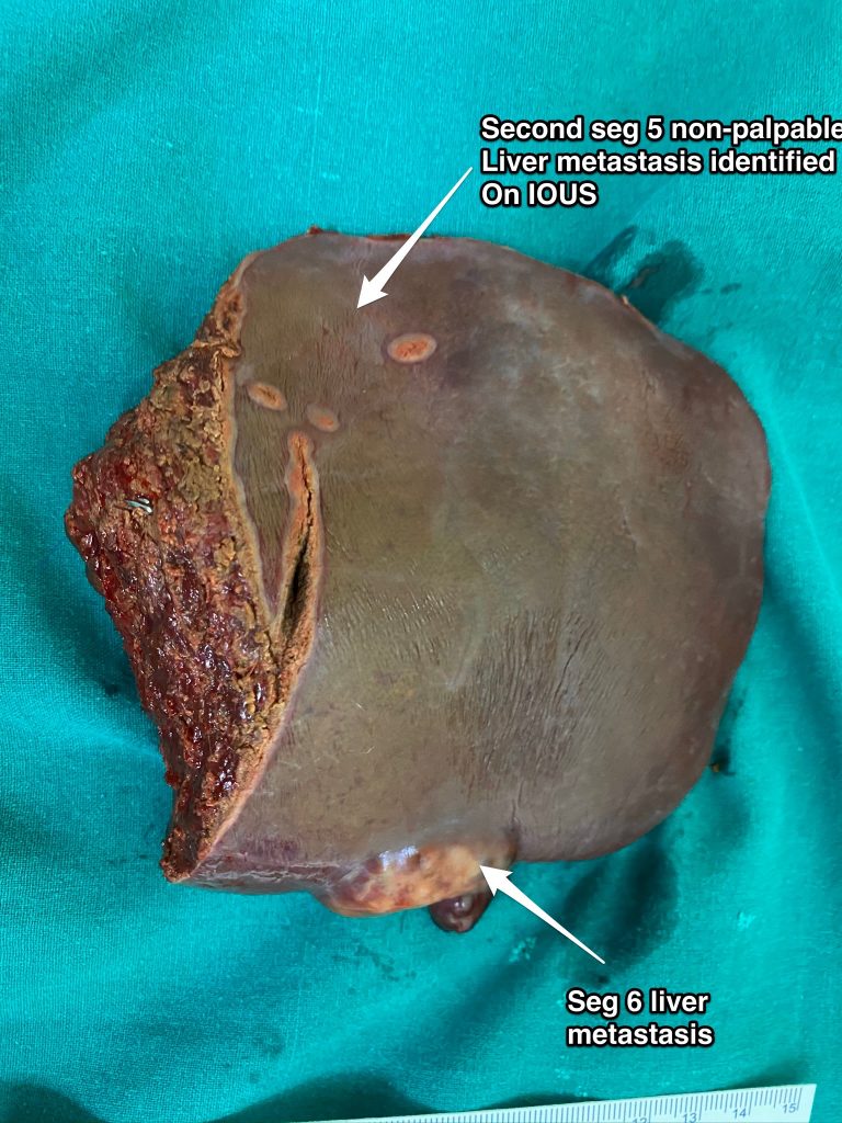
Fig 1 A

Fig 1 B

Fig. 1A and 1B – Lesion in seg 6 and another non-palpable lesion in segment 5. In the absence of IOUS, it would involve a formal set 5 resection but with IOUS, a contiguous resection can be performed with adequate margins without sacrificing additional normal liver parenchyma

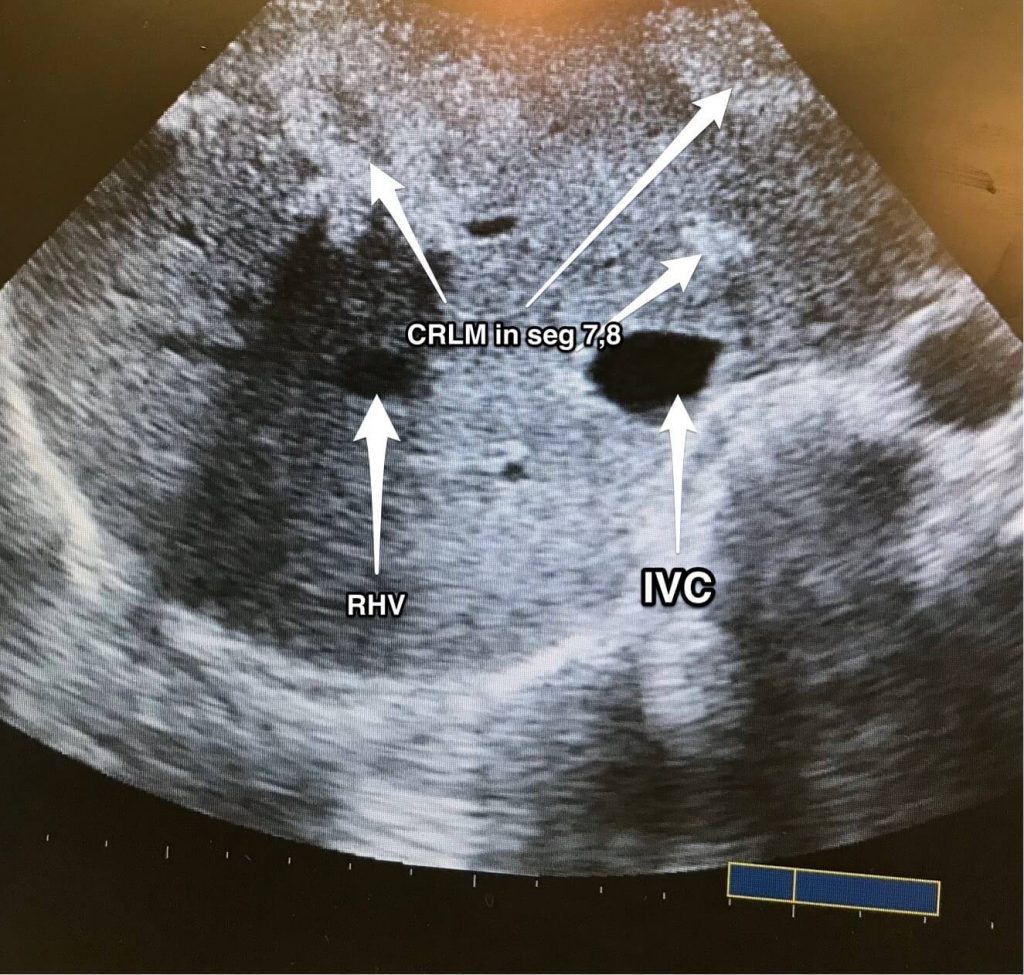
Fig 2A – IOUS identifying 3 lesions straddling segments 7,8 above the plane of the hepatic veins

Fig 2B – Resection performed under IOUS guidance to resect segments 7 and 8 above the plane of the hepatic veins to encompass the 3 liver metastases. An additional small liver metastasis identified in segment 6 was removed separately

Fig 2C – Final specimens

Fig 2 A

Fig 2 B

Fig 2 C

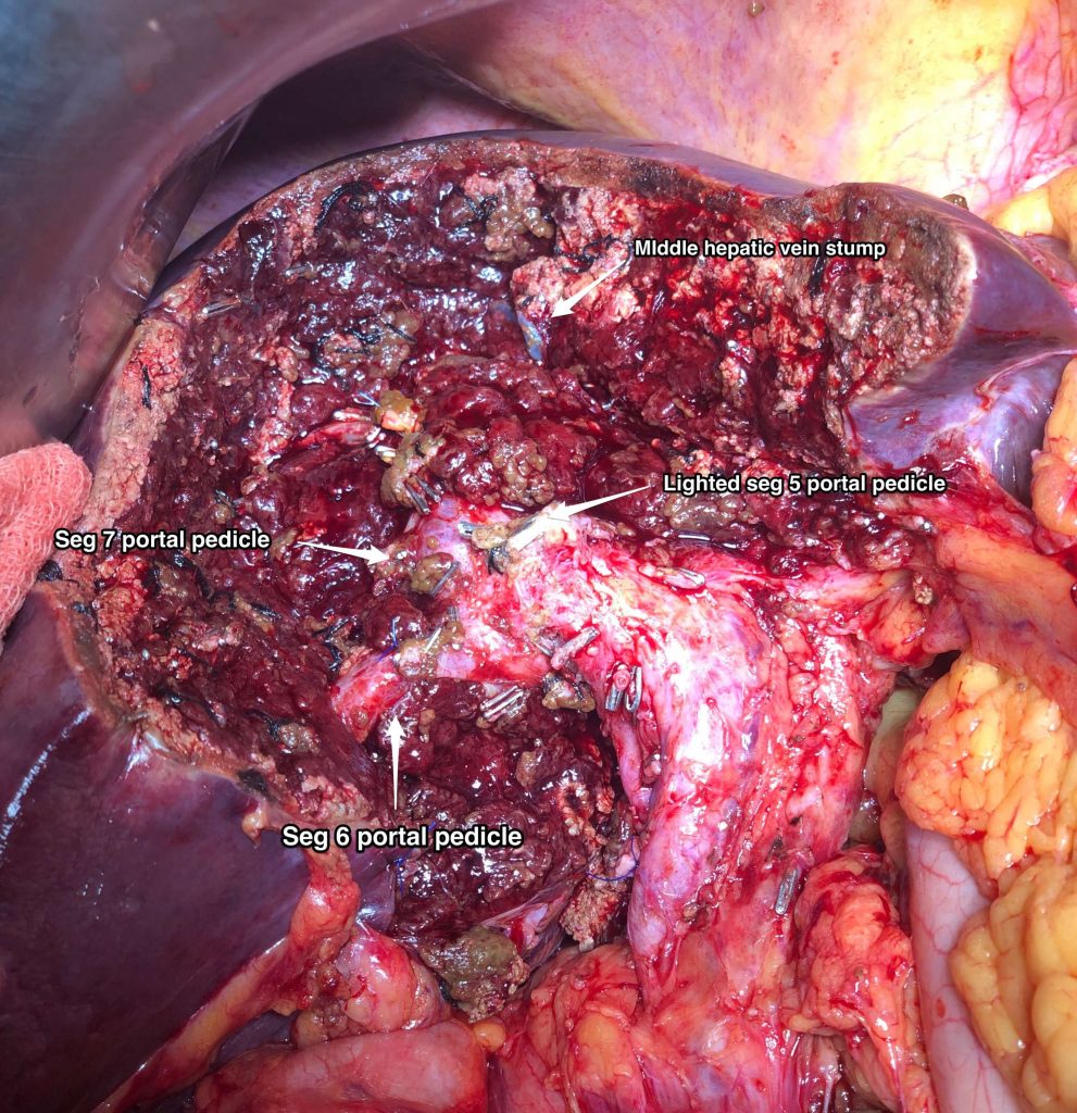
Fig 3 - A precise resection of seg 4B, 5, 8p done under IOUS guidance to resect large CRLM in close relation to the right portal pedicle

Categories
Blogs

Appendix Tumors

Appendix Tumors

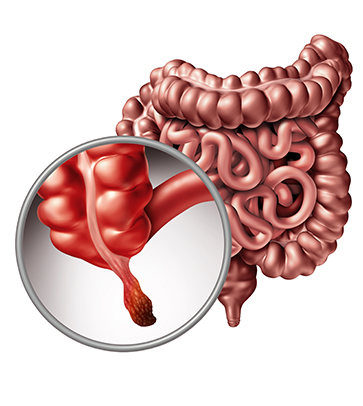
The appendix is a pouch-like tube that is attached to the cecum (the first section of the large intestine or colon). The appendix averages 10 centimeters (cm) in length and is considered part of the gastrointestinal (GI) tract. Generally thought to have no significant function in the body, the appendix may be a part of the lymphatic, exocrine, or endocrine systems.

Appendix cancer occurs when cells in the appendix become abnormal and multiply without control. These cells form a growth of tissue, called a tumor. A tumor can be benign (noncancerous) or malignant (cancerous, meaning it can spread to other parts of the body). Another name for this type of cancer is appendiceal cancer.

Types of Appendix Tumors

Carcinoid Tumor
A carcinoid tumor starts in the hormone-producing cells that are normally present in small amounts in almost every organ in the body. A carcinoid tumor arises primarily in either the GI tract or lungs, but it also may occur in the pancreas, a man’s testicles, or a woman’s ovaries. An appendix carcinoid tumor most often occurs at the tip of the appendix. Approximately 66% of all appendix tumors are carcinoid tumors. This type of cancer usually causes no symptoms until it has spread to other organs and often goes unnoticed until it is found during an examination or procedure performed for another reason. An appendix carcinoid tumor that remains confined to the area where it started has a high chance of successful treatment with surgery.
Colonic-Type Adenocarcinoma
Colonic-type adenocarcinoma accounts for about 10% of appendix tumors and usually occurs at the base of the appendix. This type of tumor looks and behaves like the most common type of colorectal cancer. It often goes unnoticed, and diagnosis is frequently made during or after surgery for appendicitis (inflammation of the appendix that can cause abdominal pain or swelling, loss of appetite, nausea, vomiting, constipation or diarrhea, inability to pass gas, or a low fever that begins after other symptoms).
Signet-Ring Cell Adenocarcinoma
Signet-ring cell adenocarcinoma (so called because, under the microscope, the cell looks like it has a signet ring inside it) is very rare and considered to be more aggressive and more difficult to treat than other types of adenocarcinomas. This type of tumor usually occurs in the stomach or colon, and it can cause appendicitis when it develops in the appendix.
Paraganglioma
Paraganglioma is a rare tumor that develops from cells of the paraganglia, a collection of cells that come from nerve tissue that persist in small deposits after fetal (pre-birth) development, and is found near the adrenal glands and some blood vessels and nerves. This type of tumor is usually considered benign and is often successfully treated with the complete surgical removal of the tumor. Paraganglioma is very rare outside of the head and neck region.
Mucinous Cystadenocarcinoma
Mucinous cystadenocarcinoma is the most common non-carcinoid appendix tumor and accounts for about 20% of appendix cancer cases. This type of tumor produces a jelly-like substance called mucin that can fill the abdominal cavity and can cause abdominal pain, bloating, and changes in bowel function if the tumor breaks through the appendix or grows in the abdomen. This is called as Pseudomyxoma Peritonei or “jelly belly”.
Appendiceal Tumors
Appendiceal tumors are often misdiagnosed as appendicitis. Alternatively, they may burst with very little symptoms and spread within the abdominal cavity to give rise to pseudomyxoma peritonei or peritoneal carcinomatosis. Fortunately, now we can treat these conditions using Cytoreductive Surgery and Hyperthermic Intra-peritoneal chemotherapy (HIPEC). Dr. Sanket Mehta has pioneered this form of treatment in the country. He has already performed numerous such surgeries with good results.